Brown Jelly Disease: Recognition and Treatment
Brown Jelly Disease: Recognition and Treatment
Brown jelly disease (BJD) is the LPS keeper’s nightmare: a healthy coral can become covered in a brown mucus mass within hours, and at worst a single polyp disappears within a day. The disease strikes fleshy LPS corals in particular — Euphylliidae (hammer, torch, frogspawn), elegance and acans are the typical victims.
It progresses fast and spreads through the water, so your response has to be fast too. The good news is that once you understand what’s going on, treatment is straightforward and the success rate is reasonable — as long as you act in time and also correct the underlying cause. This article covers what causes the disease, how to distinguish it from harmless mucus production, what to do immediately and how to dip the coral correctly.
What brown jelly is — and what causes it
As the name suggests, the disease appears as a brown, gelatinous mass on top of the coral tissue. The mass is not just mucus but a mixture of dead coral tissue, mucus, bacteria and — the decisive factor — protozoa.
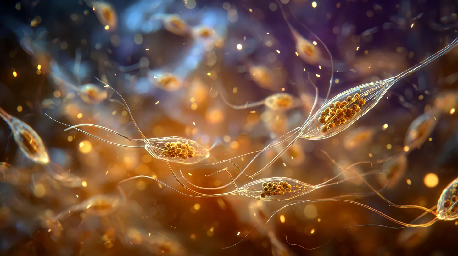
The disease was previously thought to be bacterial, but peer-reviewed research (Sweet et al. 2013) showed that the real engine of the jelly is a ciliate (a protozoan). The study identified a Philaster-genus ciliate (previously misidentified as Helicostoma notatum) that burrows into the coral tissue and ingests it. The coral’s own zooxanthellae were found inside the ciliate — it is the ingested coral tissue that gives the mass its characteristic brown colour. The same ciliate also appears in white syndrome, but in brown jelly there are far more of them, which explains the typical jelly-like form.
A key finding is that no single “guilty” bacterium was found behind it. Brown jelly is therefore not a contagious disease in the sense of a single pathogen spreading; it almost always starts from the coral weakening: poor water quality, stress, transport damage or mechanical damage that opens a route into the tissue for the ciliates and bacteria. Once the process has begun, however, the jelly and its ciliates can spread from coral to coral through the water — which is why isolation matters.
What it looks like and how fast it progresses
Brown jelly is recognised by a brown, loose, slimy mass that covers the tissue and appears to rise off it. Unlike healthy mucus, it does not rinse away in the flow but clings and grows. Beneath the mass the tissue detaches from the skeleton, and progression is fast: the loss can consume roughly 15–20 mm² of tissue per day, and a whole Euphyllia head can disappear in a day.
The most important distinction is harmless mucus production vs. disease:
- Normal mucus: many LPS corals, Euphylliidae in particular, produce clear or pale mucus under stress, after a move or as part of normal tissue cleaning. This mucus is usually translucent, rinses away in the flow, and there is no tissue detachment beneath it. The polyps extend again soon.
- Brown jelly: the mass is distinctly brown and gelatinous, it clings to the tissue, and bare skeleton is revealed beneath it. The polyp doesn’t open but shrinks under the mass.
Also distinguish the disease from bleaching (the coral pales but the tissue stays on the skeleton) and from mere tissue recession (the tissue shrinks but no brown jelly forms). If you see brown jelly and an advancing border between bare skeleton and tissue, it is almost certainly brown jelly.
Which corals are susceptible and why
Brown jelly favours fleshy, heavily mucus-producing LPS corals, whose soft tissue offers the ciliates a favourable substrate:
- Euphylliidae (hammer, torch, frogspawn) are the most typical. Wall forms in particular are more susceptible than branching forms, and a stressed or newly acquired specimen is at greatest risk.
- Elegance coral (Catalaphyllia jardinei) is notoriously disease-prone, brown jelly included.
- Acans, lobophyllia and other fleshy LPS corals can also fall ill, as can plate corals (Fungia).
One phenomenon typical of Euphylliidae corals is polyp bailout: under severe stress a polyp can detach from its skeleton entirely as a last-resort survival mechanism. A detached, healthy polyp can sometimes resettle and survive, even if the original skeleton is lost.
Deeper on this: The genus Euphyllia: deep dive
First aid — act immediately
Brown jelly progresses in hours, so speed is decisive. Act as soon as you recognise the brown jelly:
1. Isolate. Move the affected coral to quarantine or a separate container. This stops the jelly and its ciliates from spreading to other corals and gives you room to work.
2. Remove the jelly physically. This is the single most important step. Gently siphon the mass away with a turkey baster or hose — preferably outside the tank, so the detached jelly doesn’t spread into the water. Discard the contaminated water and don’t pour it back into the tank. Repeat several times until the visible mass is gone.
3. Decide whether to cut. If the coral is multi-headed (e.g. a branching Euphyllia) and some heads are still healthy, cut the healthy heads off and treat them separately. The affected part that has lost its tissue won’t recover, but clean heads can be saved.
4. Dip. Treat the coral or the cut heads with a dip (see below).
5. Fix the water quality. Because the disease almost always starts from weakening, measure and stabilise the parameters. Without correcting the underlying cause the disease returns, even if the jelly is removed for a while.
Dip options
Dipping kills or removes the ciliates and slows the disease’s progress. Use one or more of the following, and always dip outside the tank in a separate container.
Iodine dip. The gentlest and most commonly used. You can use Lugol’s solution, a commercial coral iodine, or pharmacy Betadine (10 % povidone-iodine), which works as a dip equivalently to Lugol’s and is easily available in Europe. Dose it into tank water until the water turns a light tea-brown (a few drops per decilitre), and bathe the coral in the solution for about 10–15 minutes, agitating occasionally. Iodine is gentle on the tissue and works well on the jelly. Rinse the coral in clean tank water after the dip, and don’t pour the dip water into the tank.
Hydrogen peroxide dip. More effective but harsher. Use ordinary pharmacy 3 % hydrogen peroxide (the same disinfectant sold for wound cleaning). Dilute about 1 ml / 250 ml of tank water (or 1 part peroxide to 10 parts tank water) and bathe the coral in the solution for 5–10 minutes. Don’t exceed the time — peroxide stresses the tissue. Rinse the coral in clean tank water after the dip.
The Euphyllia quirk. Euphylliidae corals pull their tentacles in under stress and shield the disease inside as they do, so the dip doesn’t work properly. Move the coral into the dip container and wait up to 30 minutes for it to open fully before adding the dip solution slowly. This way the solution reaches the parts protected by the tentacles too.
General rules: don’t put frags from different corals in the same dip container, and discard the dip water after each use — never pour it back into the tank.
More on this: Coral dipping and quarantine protocol
Fix the root cause
Removing the jelly and dipping stop the acute situation, but brown jelly returns if the coral stays weakened. Work through the underlying causes:
- Water quality and stability. Measure alkalinity, temperature, pH and nutrients. Fluctuating parameters stress the coral and predispose it to the disease. Stability is the single most important factor.
- Mechanical and transport damage. Damaged tissue is an entry point for the disease. Handle corals gently and let a new specimen recover from transport in peace.
- Overfeeding and detritus. Excess food and detritus collecting on the coral feed bacteria. Keep the flow such that debris doesn’t accumulate, and don’t overfeed.
- Stress from placement. A neighbouring coral’s burn or wrong light/flow weakens the coral. Check the placement.
More on this: Water changes in practice
Prevention
Brown jelly is easier to prevent than to treat:
- Choose a healthy specimen. Don’t buy a coral with mucus or jelly on its tissue, with receded tissue, or with skeleton showing through. A well-extended, intact coral is safest.
- Quarantine and dip. Dip every new coral and, where possible, keep it in quarantine before moving it to the display tank.
- Handle gently. Avoid damaging the tissue during moves and fragging.
- Keep conditions stable. Steady parameters and good water quality keep the coral’s defence in order.
- Observe daily. The sooner you notice the brown jelly, the more likely you are to save the coral. One early-detected, physically removed mass is far easier to deal with than the loss of a whole head.
More on this: LPS corals in practice · RTN and STN: recognising and treating tissue loss
References
Peer-reviewed studies
- Sweet, M., Craggs, J., Robson, J., & Bythell, J. (2013). Assessment of the microbial communities associated with white syndrome and brown jelly syndrome in aquarium corals. Journal of Zoo and Aquarium Research, 1(2), 67–74.
- Sweet, M., & Bythell, J. (2012). Ciliate and bacterial communities associated with White Syndrome and Brown Band Disease in reef-building corals. Environmental Microbiology, 14(8), 2184–2199.
Hobbyist literature and sources
- Tidal Gardens. Euphyllia & Fimbryphyllia Troubleshooting Guide (brown jelly, polyp bailout). Video transcript, 2024. https://tidalgardens.com/
- Manta Systems. Brown Jelly Disease in Saltwater Corals: A Complete Guide. https://www.mantasystems.net/a/blog/post/BJD
- Reef2Reef Community. Brown jelly disease — treatment strategies. https://www.reef2reef.com/
Books
- Borneman, E. H. (2001). Aquarium Corals: Selection, Husbandry, and Natural History. T.F.H. Publications.
- Delbeek, J. C., & Sprung, J. (2005). The Reef Aquarium: Science, Art, and Technology, Vol. 3. Ricordea Publishing.